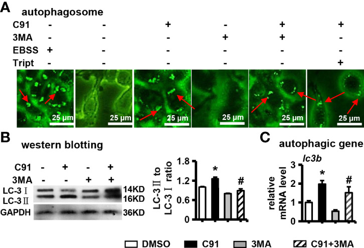
Figure 4.

The effect of C91 on autophagy in ST2 cells. (A) 3MA is an autophagy inhibitor. ST2 cells were pre-treated with 3MA (5 mM) for 6 hours, and then induced by C91 for 3 days. MDC staining was performed and autophagy was analyzed by fluorescence microscopy. Body formation. Red arrows indicate autophagosomes, scale bar = 25 μm. (B) Western blot detection of the expression of autophagy-related protein LC-3I/II. GAPDH was used as an internal control, and the histogram on the right is the quantification of the band on the left. (C) qRT-PCR analysis of the mRNA expression of autophagy-related gene lc3b. Results are expressed as mean ± SD (n = 3 per group). * indicates P < 0.05, compared with the DMSO group. # indicates P < 0.05, compared with the C91 group.
