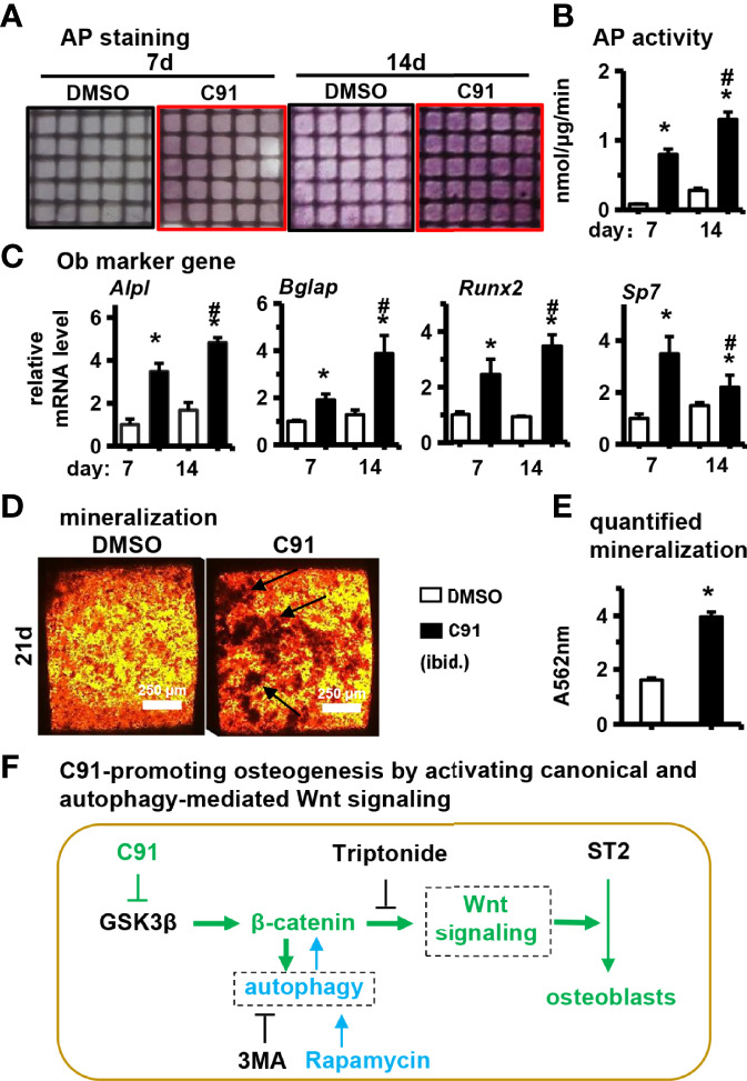
Figure 8.

Effects of C91 on ST2 cell differentiation and mineralization in 3D modules. 3D modules were cultured in complete medium for 2 days and then switched to osteogenic induction medium. The medium was changed in half every two days. After 7 and 14 days of culture, AP staining (A) analysis and AP biochemical quantification (B) analysis were performed. (C) qRT-PCR detection of the effect of C91 in the 3D module on the expression of osteogenic target genes in ST2 cells. (D) 3D modules were induced to mineralize for 21 days, followed by Alizarin Red S staining to detect mineralized nodules. Black arrows indicate mineralized nodules. Scale bar = 50 μm. (E) is the quantitative analysis result of panel (D, F) C91-promoting osteogenesis by activating canonical and autophagy-mediated Wnt signaling. *Indicates P<0.05, compared to the 7-day DMSO group. # indicates P<0.05, compared with the 14-day DMSO group.
