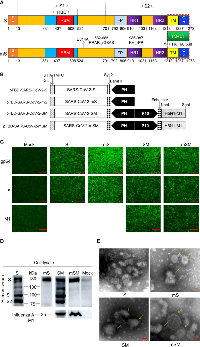
Figure 1.

Generation of SARS-CoV-2 VLPs. (A) Schematic of SARS-CoV-2 S/M1 and its mutation. (B) Schematics of the four recombinant bacmid rFBD-SARS-CoV-2. (C, D) Expression of exogenous genes by recombinant baculoviruses identified by IFA (C) and Western blot (D). The scale bar corresponds to 150 μm. Convalescent serum of COVID-19 patient or Influenza A M1 as the primary antibody, and HRP-labeled Goat Anti-Mouse IgG (H+L) as the secondary antibody. Mock, wild baculoviruses infected cell. Unprocessed original images can be found in Supplemental Figure S6. (E) Transmission electron micrograph of negatively stained SARS-CoV-2 VLPs. The scale bar corresponds to 50 nm.
