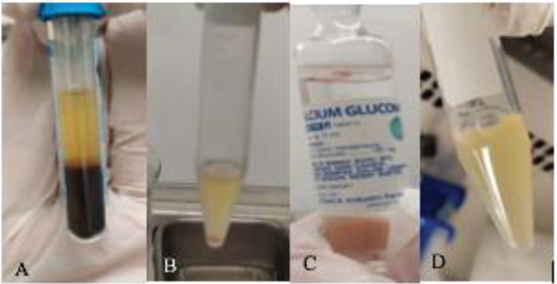
Fig. 1.

Vein cubitus blood after the first centrifugation. ( A ) shows the plasma layer, the buffy coat layer, and the PRP layer (which formed after the second centrifugation). ( B ) shows the addition of 10% calcium gluconate in the PRP-T tube. ( C ) shows the gel phase that forms 15 minutes after the procedure (at 37°C). PRP-T, platelet rich plasma thrombin.
