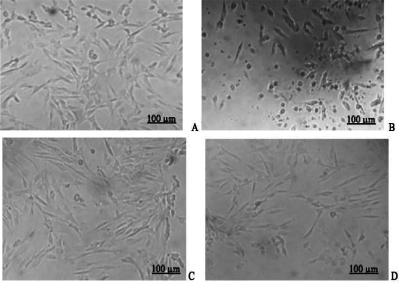
Fig. 2.

The hDPSCs morphology before ( A ) and after ( B ) starvation; hDPSCs in different cultured media; and PRP ( C ) and PRP-T groups ( D ) after 24 hours of cultured (scale of 100 μm) (Inverted microscope, Zeiss, Observer Z1, UK). hDPSC, human dental pulp stem cell; PRP-T, platelet rich plasma thrombin.
