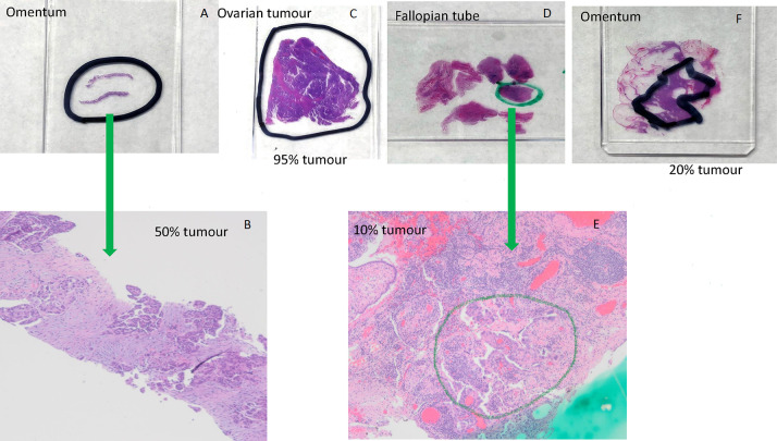
Figure 1.
Examples of marking tumour area for dissection and estimation percent of tumour using H&E-stained sections. (A) Omentum; (B) omentum, 40× magnification, 50% tumour cellularity. High-grade serous carcinoma with solid nests and papillary-like clusters of malignant cells within a reactive fibroblastic stroma; (C) ovarian tumour, 95% tumour cellularity. Almost entirely high-grade serous carcinoma with papillary structures and slit-like spaces, with a small focus of background non-neoplastic fibrous tissue; (D) fallopian tube; (E) fallopian tube, 40× magnification, 10% tumour cellularity. Minute focus of residual high-grade serous carcinoma postinterval neoadjuvant chemotherapy, rimming papillary stromal cores. Approximately 20% cellularity in the circled area, within a background of reactive fibroblastic proliferation and chronic inflammatory cells; (F) omentum, 20% tumour cellularity.

