
Fig. 1.
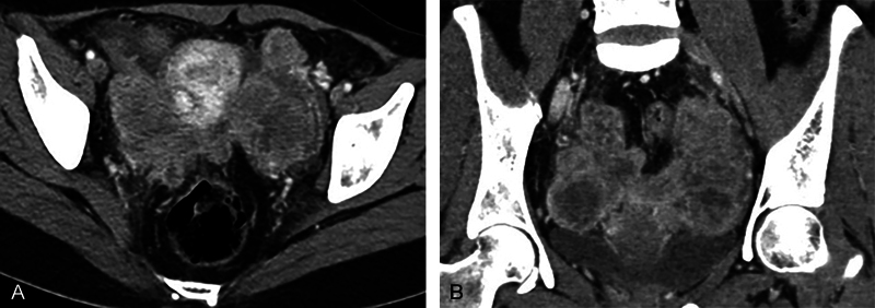
( A and B ) Axial and coronal contrast-enhanced computed tomography images show bilateral solid heterogeneously enhancing adnexal masses. Ovaries are not seen separately. Masses are closely abutting the uterus on either side.
Official websites use .gov
A
.gov website belongs to an official
government organization in the United States.
Secure .gov websites use HTTPS
A lock (
) or https:// means you've safely
connected to the .gov website. Share sensitive
information only on official, secure websites.
( A and B ) Axial and coronal contrast-enhanced computed tomography images show bilateral solid heterogeneously enhancing adnexal masses. Ovaries are not seen separately. Masses are closely abutting the uterus on either side.