Figure 3.
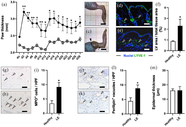
Animal model validation. LE evaluation over 28 days by means of paw thickness measurements ((a) black dots = LE, white dots = healthy). Representative stereomicroscopic images of healthy (b) and LE (c) hindlimbs on day 28 with persistent swelling (arrow in (c)) of the operated limb (arrowhead = lymphadenectomy scar). LYVE-1+ dermal lymphatic vessels (arrowheads) on day 28 in healthy (d) and LE (e) hindlimbs. Quantification of lymphatic vessel area per total tissue area in % (f). Infiltration of MPO+ neutrophilic granulocytes (arrowheads) in healthy (g) and LE (h) hindlimbs. Quantification of MPO+ cells per HPF (i). Perilipin+ fat vacuoles (arrowheads) in healthy (j) and LE (k) hindlimbs. Quantification of perilipin+ vacuoles per HPF (l). Quantification of epidermal thickness in µm (m). (a, f, i, l, and m) Mean ± SEM, n = 8, *p < 0.05 versus healthy, **p < 0.001 versus healthy. Scale bars: (b) and (c) = 5 mm, (d, e, g, h, j, and k) = 60 µm. HPF: high-power field; LE: lymphedema; LV: lymphatic vessel; LYVE: lymphatic vessel endothelial hyaluronan receptor; MPO: myeloperoxidase.

