Figure 5.
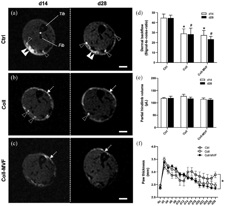
Volumetry and dermal backflow after MVF transplantation. Axial MR lymphography scans of control (a), collagen (b), and collagen/MVF (c) LE hindlimbs at the level of the distal tibio-fibular joint (asterisk) 14 and 28 days after lymphadenectomy. Dermal backflow ((a) double arrowheads) is observed in all groups with a significant reduction in the collagen and collagen/MVF groups ((b and c) dashed arrows). Dashed arrowheads: individual lymphatic vessels, Fib: fibula; Tib: tibia. Quantification of dermal backflow (d). *p < 0.05 versus Ctrl day 14, #p < 0.05 versus Ctrl day 28. Mean ± SEM, n = 8. Quantification of hindlimb volumes by means of MR volumetry (e) and paw thickness measurements (f). *p < 0.05 versus Ctrl. Mean ± SEM, n = 8–9. Scale bars = 1 mm. Coll: collagen; Ctrl: control; LE: lymphedema; MR: magnetic resonance; MVF: microvascular fragments.

