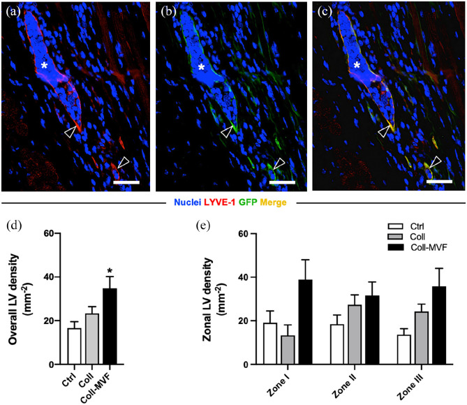
Figure 7.
Popliteal lymphatic vessel density after MVF transplantation. Immunohistochemical detection (a–c) of MVF-derived LYVE-1+/GFP+ lymphatic vessels (arrowheads). Scale bars = 30 µm. Quantification of overall LV density in mm−2 (d). Quantification of zonal LV density in mm−2 (e). Mean ± SEM, n = 8–9, *p < 0.05 versus Ctrl. Coll: collagen; Ctrl: control; GFP: green fluorescent protein; LV: lymphatic vessel; LYVE: lymphatic vessel endothelial hyaluronan receptor; MVF: microvascular fragments.

