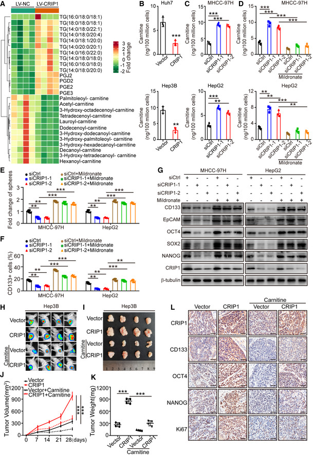
Figure 3. CRIP1 downregulates carnitine to facilitate stemness in HCC.

-
ALipidomics analysis of CRIP1‐overexpressing Huh7 cells.
-
BCarnitine levels in CRIP1‐overexpressing Huh7 and Hep3B cells.
-
CCarnitine levels in CRIP1 knockdown MHCC‐97H and HepG2 cells.
-
DCarnitine levels in CRIP1 knockdown MHCC‐97H and HepG2 cells with or without 5 mM mildronate for 24 h.
-
ESphere formation ability in the indicated groups.
-
FFlow cytometric analysis revealed the proportions of CD133+ cells in the indicated groups.
-
GWestern blot analysis of cancer stem cell markers in the indicated groups.
-
H, IIn vivo bioluminescent images (H) and bright‐field images (I) of xenografts in mice with the indicated Hep3B cells. Carnitine was added to drinking water to a final concentration of 4.0 mg/ml.
-
J, KThe growth curve of tumors (J) and quantification of tumor weight (K) are presented (n = 4).
-
LIHC analysis of CRIP1 and HCC cancer stem cell markers. Scale bar: 20 μm.
Data information: Data represent the mean ± SD at least three independent experiments. **P < 0.01 and ***P < 0.001. Differences were tested using an unpaired two‐tailed Student's t‐test (B–F, J, K).
