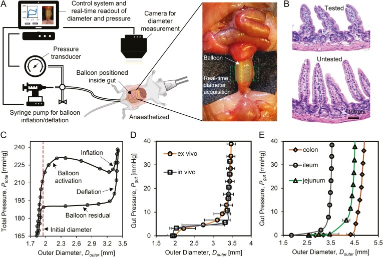
Figure 1.
Schematic of real-time gut diameter and stiffness measurement using mechanoscope. A, The mechanoscope includes control system, pressure transducer, balloon catheter, syringe pump for the balloon inflation/deflation, and gut diameter measurement camera. The control system coordinates the pressure transducer and gut diameter measurement camera to automatically record real-time pressure and diameter. The gut diameter measurement module is integrated into the control system, which identifies the gut (green dashed box) and detects the gut edges (green solid line). B, H&E staining shows that during testing, no damage of mucosa and muscle layer occurs in tested samples compared with an adjacent untested section of ileum. C, In testing cycles the gut outer diameter, Douter is recorded in correspondence with total pressure, Ptotal. A test cycle includes 4 phases, balloon activation, inflation, deflation, and balloon residual. The initial diameter (red dashed line) marks the gut diameter at rest prior to testing. D, Gut pressure (Pgut) is calculated after removing balloon residual from Ptotal. No significant differences are detected for Pgut-Douter tissue response under in vivo and ex vivo testing conditions. E, Pgut as a function of Douter are measured from different gut segmentations, jejunum, ileum, and colon samples.

