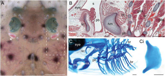
Fig. 1.
Anatomical overview of the spiracle, pseudobranch and spiracular cartilage in the little skate. (A) Dorsal view of the eye and spiracle of a skate hatchling. The spiracles are indicated with asterisks. Dashed box indicates the regions shown in B and C. (B) Frontal section of a S32 skate embryo stained with Masson's trichrome showing the spiracular region. The pseudobranch sits inside the spiracle and is supported by the spiracular cartilage in the anterior spiracle wall. (C) Lateral view of a S32 skate embryo skeletal preparation stained with Alcian Blue showing the pharyngeal endoskeleton. The spiracular cartilage is located dorsal to the articulation between the upper jaw (palatoquadrate) and the lower jaw (Meckel's cartilage). (Ci) A dissected skate spiracular cartilage. The leaf-like sheet of cartilage supports the anterior wall of the spiracle. br, branchial rays; eb, epibranchials; hm, hyomandibula; mk, Meckel's cartilage; pb, pseudobranch; ph, pseudohyal; pq, palatoquadrate; s, spiracle; sc, spiracular cartilage. Scale bars: 3 cm in A; 500 μm in B-Ci.

