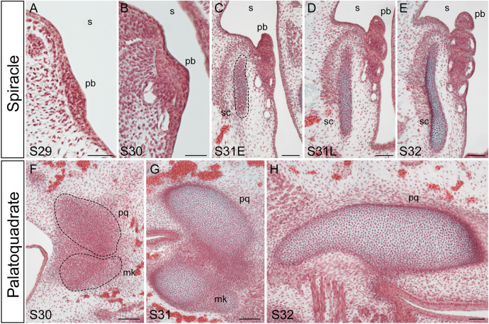
Fig. 2.
Development of the pseudobranch, spiracular cartilage and palatoquadrate in the little skate. Frontal sections stained with modified Masson's trichrome. (A) At S29, the development of the pseudobranch is first apparent as a thickening of the posterior epithelium of the mandibular arch. (B) By S30, the pseudobranch thickens further and vasculature lies adjacent to it. (C,D) At S31, the pseudobranch extends from the posterior epithelium of the mandibular arch and takes on a lamellar-like morphology; the spiracular cartilage is condensing within subjacent mesenchyme (dashed line in C). (E) By S32, the pseudobranch and spiracular cartilage have fully differentiated. (F-H) The spiracular cartilage chondrifies completely independently of the condensing (F) and differentiating (G,H) palatoquadrate. mk, Meckel's cartilage; pb, pseudobranch; pq, palatoquadrate; s, spiracle; sc, spiracular cartilage. Scale bars: 50 μm.

