Fig. 7.
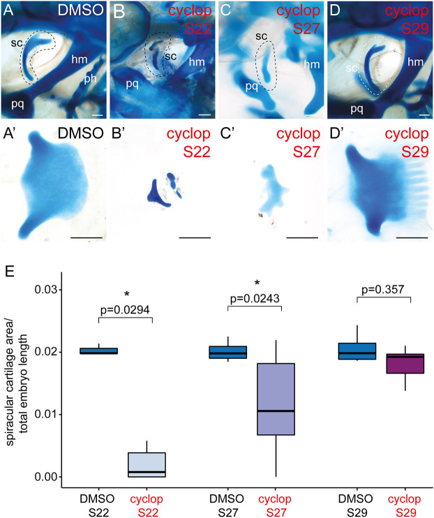
Patterning of the skate spiracular cartilage by shh signalling. (A,C′) DMSO (control) treatment in ovo has no effect on spiracular cartilage morphology (A,A′), while treatment with 20 µM cyclopamine in ovo at S22 (B,B′) and S27 (C,C′) causes spiracular cartilage malformation and a reduction in spiracular cartilage size. (D,D′) Treatment with 20 µM cyclopamine in ovo at S29 has no significant effect on spiracular cartilage size. (E) Analysis of spiracular cartilage size (spiracular cartilage area/total embryo length) between cyclopamine- and DMSO-treated skate embryos at S22, S27 and S29 reveals statistically significant reductions in spiracular cartilage size with cyclopamine treatment at S22 and S27, but no significant reduction in spiracular cartilage size with cyclopamine treatment at S29. Kruskal–Wallis test followed by Dunn's test with Bonferroni correction. The horizontal lines represent the median, the boxes indicate the 1st and 3rd quantiles, and the whiskers indicate the maximum and minimum data points. hm, hyomandibula; ph, pseudohyal; pq, palatoquadrate; sc, spiracular cartilage. Scale bars: 500 μm.

