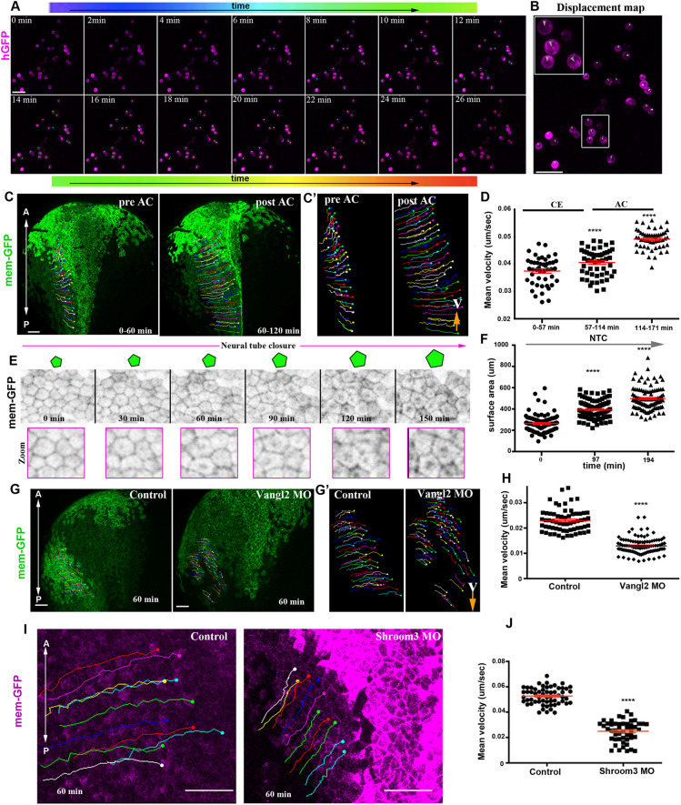
Fig. 5.
SE medial movement requires forces generated by NP morphogenesis. (A) Stills from a time-lapse recording of deep SE cells plated on an FN-coated coverslip. Tracks (spots) are time colour-coded. (B) Displacement map from a 26 min time window from the time-lapse recording used for cell tracking in A, indicating absence of cell movement. Boxed area is shown at higher magnification in inset. (C) Stills from a tracked time-lapse recording of a control embryo before (left) and after (right) AC. (C′) Zoomed tracks from the recording shown C showing the increase of cell displacement and velocity (V) during AC. (D) Quantification of the average cell velocity of SE cells during distinct phases of NTC. ****P<0.0001 (two-sided, unpaired Student's t-test); mean±s.e.m. n=46, 53 and 54 cells for the three different time periods. (E) Stills from a tracked time-lapse recording showing SE cells during NTC. As NTC progresses, the surface area of SE cells increases, as shown by the schematics above. (F) Quantification of the apical surface area of SE cells during NTC. ****P<0.0001 (two-sided, unpaired Student's t-test); mean±s.e.m. n=100 cells for each time point. (G) Stills from a tracked time-lapse recording of a control embryo (left) and a Vangl2 morphant embryo (right). (G′) Zoomed tracks from the recording shown in C showing decrease of cell displacement and velocity (V) in the absence of CE in Vangl2 morphants. (H) Quantification of the average cell velocity from control and Vangl2 morphant embryos. ****P<0.0001 (two-sided, unpaired Student's t-test); mean±s.e.m., n=63 SE cells from a control and 97 SE cells from a Vangl2 morphant embryo. (I) Stills from a tracked time-lapse recording of a control embryo (left) and a Shroom3 morphant embryo (right). The displacement and velocity (V) of SE cells is decreased in the absence of AC. (J) Quantification of the average cell velocity from control and Shroom3 morphant embryos. ****P<0.0001 (two-sided, unpaired Student's t-test); mean±s.e.m., n=59 SE cells from a control and 49 SE cells from a Shroom3 morphant embryo. Scale bars: 100 µm. A, anterior; P, posterior.

