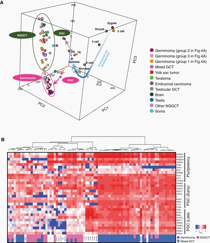
Fig. 3.
Transcriptomic comparison between normal embryonic cells at various developmental stages and GCTs of all histopathological variants. (A) Three-dimensional principal component analysis showed that primordial germ cells (PGCs) and embryonic stem cells (ESCs) diverged from the morulae/epiblast. Germinomas (circled in pink) were located further away in the direction of PGCs and nongerminomatous GCTs (circled in dark green) were situated close to ESCs. (B) Supervised hierarchical clustering of the same set of embryonic cells and the current GCT cases using PGC (early and late)-related, and pluripotency-related genes as described in a previous report.15 Germinomas were mostly clustered with PGC, nongerminomatous GCT with ESC, and the normal embryonic cells from zygote to morulae were clustered separately. Epi, epiblast; PE, primitive endoderm; PGCLC, primordial germ cell-like cell; TE, trophectoderm; Tcam2, testicular germ cell tumor cell line.

