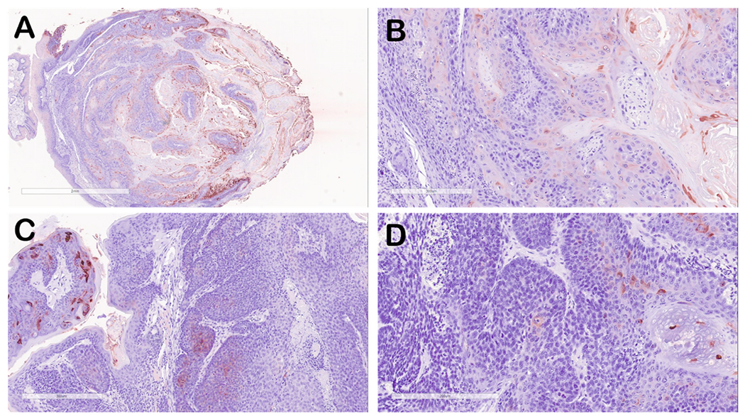
Fig. 2. Immunohistochemical staining of representative oral tumor tissues stained for MmuPV1 E4 protein (red-brown signal).

(A) lower magnification (4X) of tumor from mouse 8-2 UOM, Group 5). (B) E4 staining of tumor from mouse 8–2 (20X) showing areas of E4 staining representing areas of benign papilloma as well as tissue that is negative for E4 staining. (C) E4 staining of tumor tissue from mouse 1–3 (Group 1) showing a region of strongly stained benign papilloma (left) and tumor tissue showing minimal E4 protein. (D) E4 staining of tumor tissue from mouse 10–1 (UOM, Group 5) showing minimal E4 protein staining.
