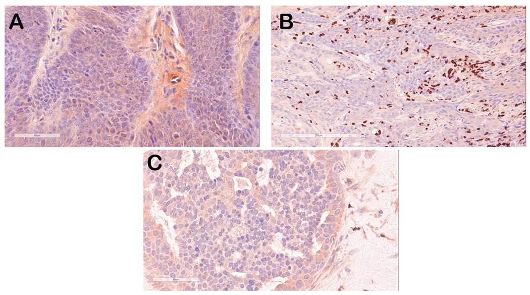
Fig. 6.

Immunostaining of mouse oral tumor tissue for S100A9 (red-brown signal). Selected mouse oral tumor tissues (A, B) were stained with anti-S100A9 antisera and showed weak to background cytoplasmic staining in tumor epithelial tissue (A) but strong staining of infiltrating neutrophils/monocytes in some tumor tissues (B), a finding that we have observed previously in some of the MmuPV1-infected mouse tissues [20]. Human breast cancer tissues also showed weak cytoplasmic staining of epithelium (C).
