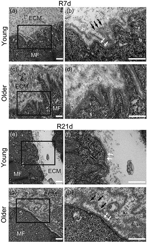
Fig. 2.

Electron microscopy analysis. Longitudinal sections of tibialis anterior (TA) muscle were observed using transmission electron microscopy (three samples per group). Basement membrane (BM) structures of young rats (a and b) and older rats (c and d) on R7d. BM structures of young (e and f) and older (g and h) rats on R21d. MF, muscle fiber; ECM, extracellular matrix; black arrow, pleated BM-like structure; white arrow, lamina densa. (b), (d), (f) and (h) are enlargements of the black squares in (a), (c), (e) and (g). Scale bar is 500 nm.
