Figure 1:

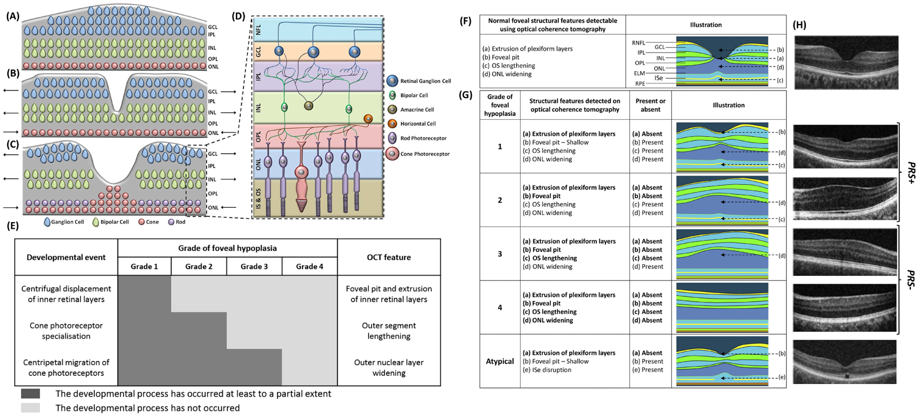
Foveal pit formation and movement of retinal cells during formation of the area of high acuity. The laminar retinal structure prior to foveal pit formation is shown in (A). The inner retinal layers were displaced centrifugally (away from the future fovea) during foveal pit formation (B). The cone photoreceptors migrate centripetally (towards the fovea) and form the pure cone area (C). Arrows point in the direction of movement of the cellular layers. The magnified laminar structure (D) with the different retinal cell types and the inner segment & outer segment (IS & OS) of the photoreceptors are shown in (D). NFL = Nerve Fibre Layer; GCL = ganglion cell layer; IPL = inner plexiform layer; INL = inner nuclear layer; OPL = outer plexiform layer; ONL = outer nuclear layer. (A, B, C are based on developmental theory proposed by Springer and Hendrickson).39 Chart showing the 3 developmental processes involved in formation of a structural and functional fovea. In grade 1 foveal hypoplasia, all processes occur to a certain extent. However, in grade 4 foveal hypoplasia, none of these processes occur; thus, the retina resembles that of the parafovea. In grade 2 and 3 foveal hypoplasia, there is outer nuclear layer widening, but no foveal pit. The difference between grade 2 and 3 foveal hypoplasia is occurrence of cone photoreceptor specialization. Identifying these specific features on optical coherence tomography (OCT) enables us to understand whether the respective developmental process has occurred. F, Illustration showing the unique features of a normal fovea detectable on optical coherence tomography. G, Illustration of typical and atypical grades of foveal hypoplasia. All grades of foveal hypoplasia had incursion of inner retinal layers. Atypical foveal hypoplasia also had incursion of the inner retinal layers. Grade 1 foveal hypoplasia is associated with a shallow foveal pit, outer nuclear layer (ONL) widening, and outer segment (OS) lengthening relative to the parafoveal ONL and OS length, respectively. In Grade 2 foveal hypoplasia, all features of grade 1 are present except the presence of a foveal pit. Grade 3 foveal hypoplasia consists of all features of grade 2 foveal hypoplasia except the widening of the cone outer segment. Grade 4 foveal hypoplasia represents all the features seen in grade 3 except there is no widening of the ONL at the fovea. Finally, an atypical form of foveal hypoplasia also is described in which there is a shallower pit with disruption of the inner segment ellipsoid (ISe). (Adapted with permission from Thomas et al. 2011).1 H, original OCTs demonstrating the different grades of foveal hypoplasia. Grades 1 and 2 can be considered to show signs of photoreceptor specialisation (PRS+), however grades 3 and 4 do not show signs of photoreceptor specialisation (PRS−). ELM = external limiting membrane; GCL = ganglion cell layer; INL = inner nuclear layer; IPL = inner plexiform layer; OPL = outer plexiform layer; RNFL = retinal nerve fibre layer; RPE = retinal pigment epithelium.
