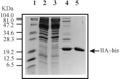
FIG. 3.
Purification of IIALacS-6H. Shown is a Coomassie brilliant blue-stained SDS–15% polyacrylamide gel. Lane 1, protein marker; lane 2, cell extract of E. coli DH5α/pSKIIAhis (50 μg of protein); lane 3, cytosolic fraction of E. coli DH5α/pSKIIAhis (50 μg of protein); lane 4, IIALacS-6H after nickel chelate affinity chromatography (∼10 μg of protein); lane 5, IIALacS-6H after nickel chelate and anion-exchange chromatography (∼7 μg of protein).

