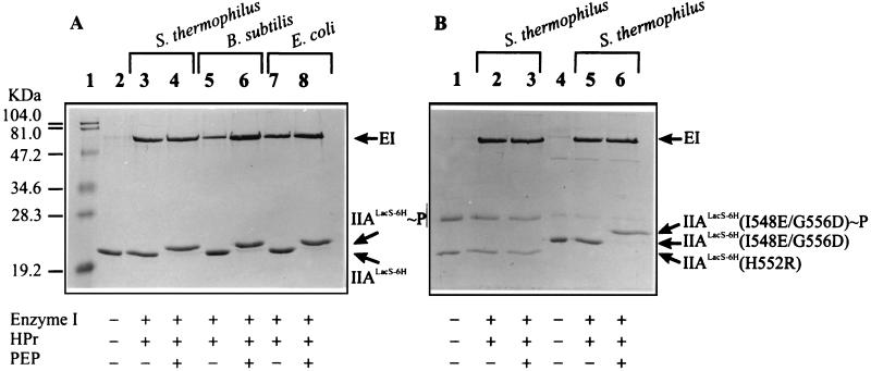
FIG. 4.
Phosphorylation of IIALacS-6H, IIALacS-6H(H552R), and IIALacS-6H(I548E/G556D). The Coomassie brilliant blue-stained SDS–15% polyacrylamide gel represents samples containing 0.8 μM enzyme I (EI) from B. subtilis, 12.5 μM HPr, 5.8 μM IIALacS-6H, and/or 10 μM PEP, as indicated at the bottom. The phosphorylation reactions (at 37°C for 15 min) were carried out in 50 mM Tris-acetate (pH 7.5)–1 mM DTT–2 mM MgCl2. (A) IIALacS. (B) Lanes 1 to 3, IIALacS(H552R); lanes 4 to 6, IIALacS-6H(I548E/G556D). The sources of HPr (S. thermophilus, B. subtilis, and E. coli) are indicated above the lanes.

