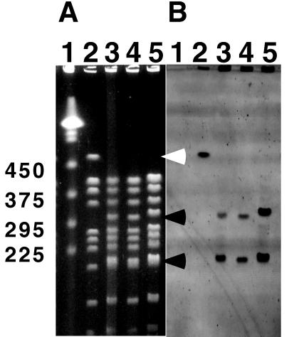
FIG. 1.
(A) PFGE analysis of wild type and transformants. DNA agarose plugs were digested with NotI restriction enzyme, and digested fragments were resolved with a contour-clamped homogeneous electric field mapper. The gel was stained with ethidium bromide. Lane 1, yeast markers; lane 2, R1 (wild type); lane 3, KKW7001 (complete katA); lane 4, KKW7002 (katA with its promoter region deleted); lane 5, KKW7003 (partial internal coding region of katA). The 485-kb NotI fragment (which disappears) is indicated by the white arrowhead. The two new NotI fragments are indicated by black arrowheads. The molecular sizes in kilobases are shown on the left. (B) Southern blot from gel shown in panel A and hybridized with fluorecein-labelled probe generated from pKKW1 plasmid DNA. The hybridization signal was detected with a FluorImager SI as described previously (24).

