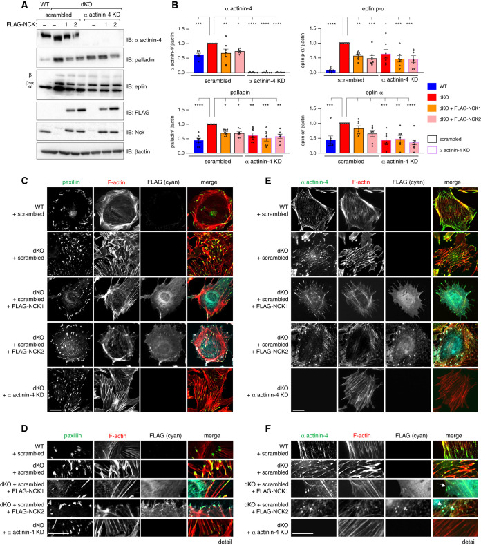
Figure 4.
Nck governs α actinin-4–directed actin bundling at stress fibers and focal adhesions. Comparison of protein levels by (A) immunoblot (IB) and (B) densitometric analysis in Nck1/2 dKO cells subjected to scrambled/nontargeting treatment (red bars, black outline) with α actinin-4 knockdown (KD) cells (red bars, purple outline) demonstrates normalization of palladin and eplin protein levels with depletion of α actinin-4 (relative to levels in WT cells [blue bars]). Similar results were achieved in dKO cells rescued with FLAG-NCK1 (orange bars) or FLAG-NCK2 (pink bars). P<0.05, **P<0.01, ***P<0.001, ****P<0.0001, two-way ANOVA and Tukey multiple comparisons test. (C–F) Immunofluorescence of WT (+scrambled) and dKO (+scrambled or α actinin-4 KD) cells with indicated rescues (FLAG-NCK1 or FLAG-NCK2). Arrows demark punctae containing F-actin, Nck, and α actinin-4. Scale bar, 100 μm.

