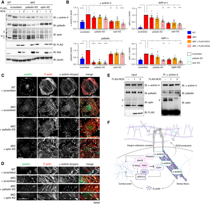
Figure 5.
Eplin and palladin differentially modulate α actinin-4. Comparison of protein levels (A) by immunoblot (IB) and (B) densitometric analysis in Nck1/2 dKO cells subjected to scrambled treatment (red bars, black outline) with palladin knockdown (KD) cells (red bars, blue outline) demonstrates normalization of α actinin-4 levels only in the presence of FLAG-NCK2 (pink bar, blue outline). Conversely, comparison of protein levels in dKO plus eplin KD cells (red bars, green outline) with dKO scrambled cells demonstrates modest normalization of α actinin-4 levels, but not those of palladin. This is independent of re-expression of FLAG-NCK1 (orange bar, green outline) or FLAG-NCK2 (pink bar, green outline). *P<0.05, **P<0.01, ***P<0.001, ****P<0.0001, two-way ANOVA and Tukey multiple comparisons test. (C–D) Immunofluorescence imaging of WT (+scrambled) and dKO (+scrambled or +palladin KD, or +eplin KD) cells. Scale bar, 100 μm. (E) Co-immunoprecipitation (IP) and IB of α actinin-4 and palladin or eplin in WT cells overexpressing FLAG-NCK1, FLAG-NCK2, or FLAG control. (F) Graphic summary of Nck-mediated regulation of the α actinin-4/β1-integrin module within the podocyte.

