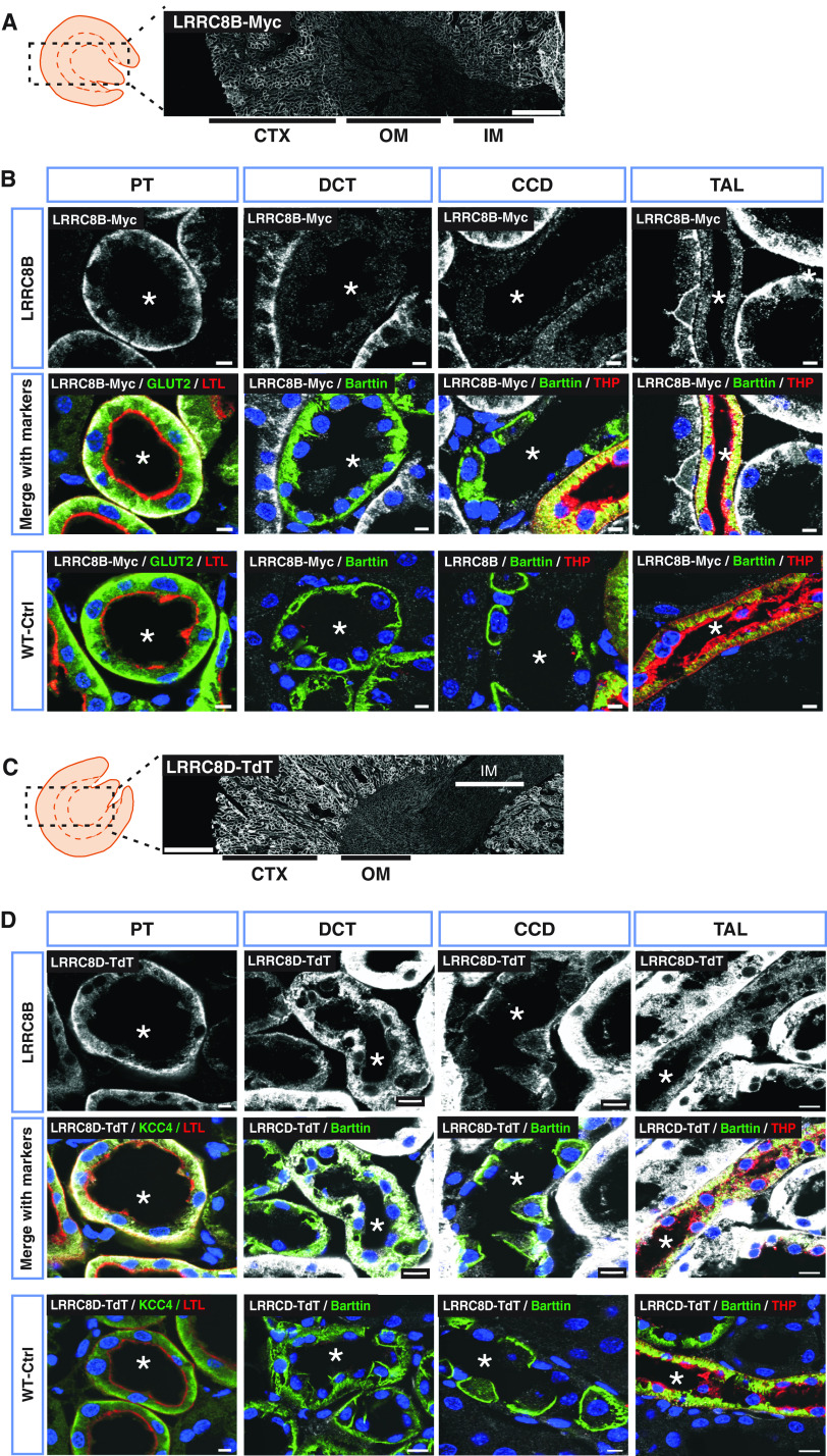
Figure 2.
Prominent expression of LRRC8B and LRRC8D in basolateral membranes of PTs. (A) LRRC8B-smFPMyc detection (white) in Lrrc8bsmFPMyclox/smFPMyclox kidney, using a Myc antibody. (B) Immunodetection of LRRC8B-smFPMyc (in white) in different renal tubules. LRRC8B-smFPMyc is prominently found at the basolateral membrane of the PT with weaker expression in the TAL, but was not detected in the DCT and CCD. (C) Overview of renal LRRC8D-TdTomato expression in Lrrc8dTdTlox/TdTlox mice, detected with an RFP antibody (white). (D) Localization of LRRC8D-TdTomato (white) in different nephron segments. In (A) and (C), kidney cortex (CTX), outer medulla (OM), and inner medulla (IM) are indicated. In (B) and (D), an asterisk indicates lumen of the tubule. PT was identified with Lotus tetragonolobus Lectin (LTL) at the brush border (red) and GLUT2 or KCC4 (green) at the basolateral membrane. DCT recognized by the basolateral staining of Barttin (green), which also expresses in ICs of CCD and TAL. TAL was additionally labeled with Tamm–Horsfall protein (THP, red) at the apical membrane. Myc and RFP-specific signal was tested in WT mice (WT-Ctrl). Nuclei stained with DAPI (blue). Scale bars, 10 μm (DCT, TAL), 5 μm (CCD, PT), and 500 μm (whole kidney). Images are representative of n≥3 mice per genotype.

