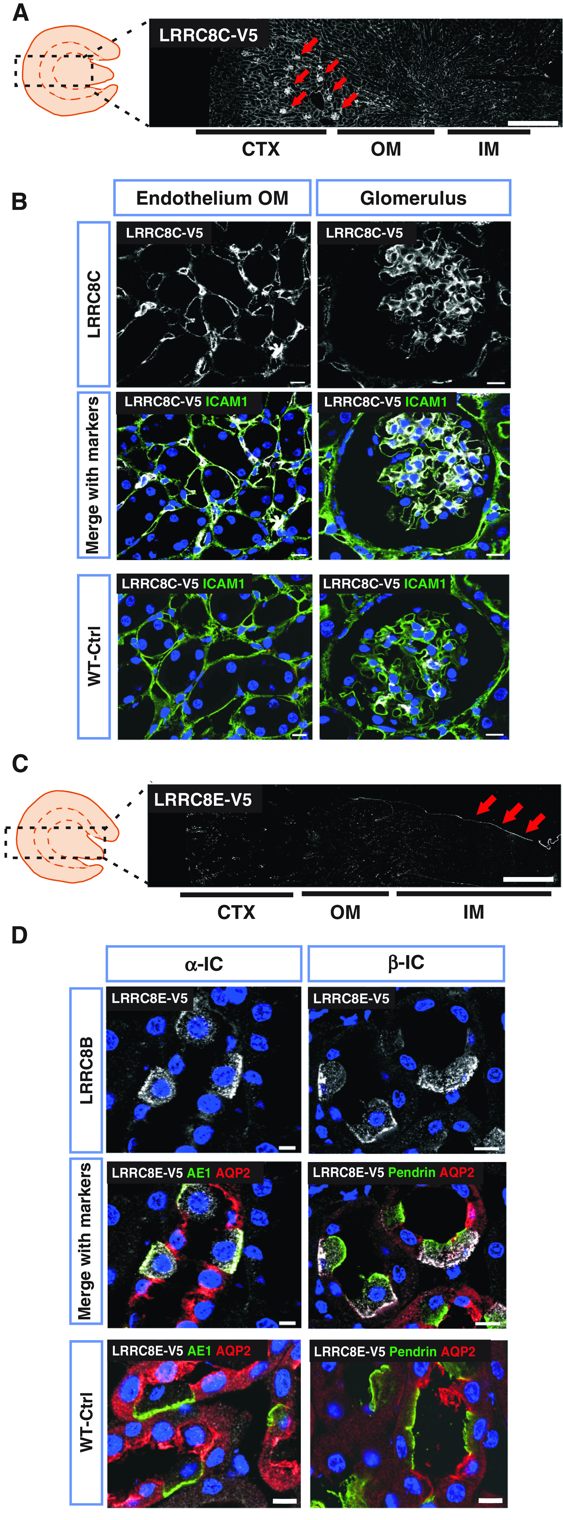
Figure 3.

Renal expression pattern of LRRC8C and LRRC8E. (A) V5-tagged LRRC8C (white) in knockin Lrrc8cV5/V5 mouse, using a V5 antibody. Glomerulus are indicated with arrows. (B) LRRC8C-V5 (in white) colocalized with the endothelial marker ICAM1 (green) in outer medulla (OM) and glomerulus. (C) Renal expression of LRRC8E-smFPV5 in Lrrc8esmFPV5/smFPV5 knockin mice, using a V5 antibody. Prominent urothelium staining (arrows), and scattered staining in medulla and cortex which represent ICs as seen in (D). (D) LRRC8E-V5 (in white) is expressed in basolateral membranes of ICs, identified as α-IC by AE1 (green) and β-IC by pendrin (green). LRRC8E-V5 was not found in principal cells (stained with AQP2 in red). In (A) and (B), cortex (CTX), outer medulla (OM), and inner medulla (IM) are indicated below. V5 signal specificity was tested in WT mice (WT-Ctrl), nuclei stained with DAPI (blue). Scale bars, 10 μm (glomerulus, DCT, PT), 5 μm (CCD, TAL), and 500 μm (whole kidney). Images are representative of at least three mice per genotype.
