Figure 7.

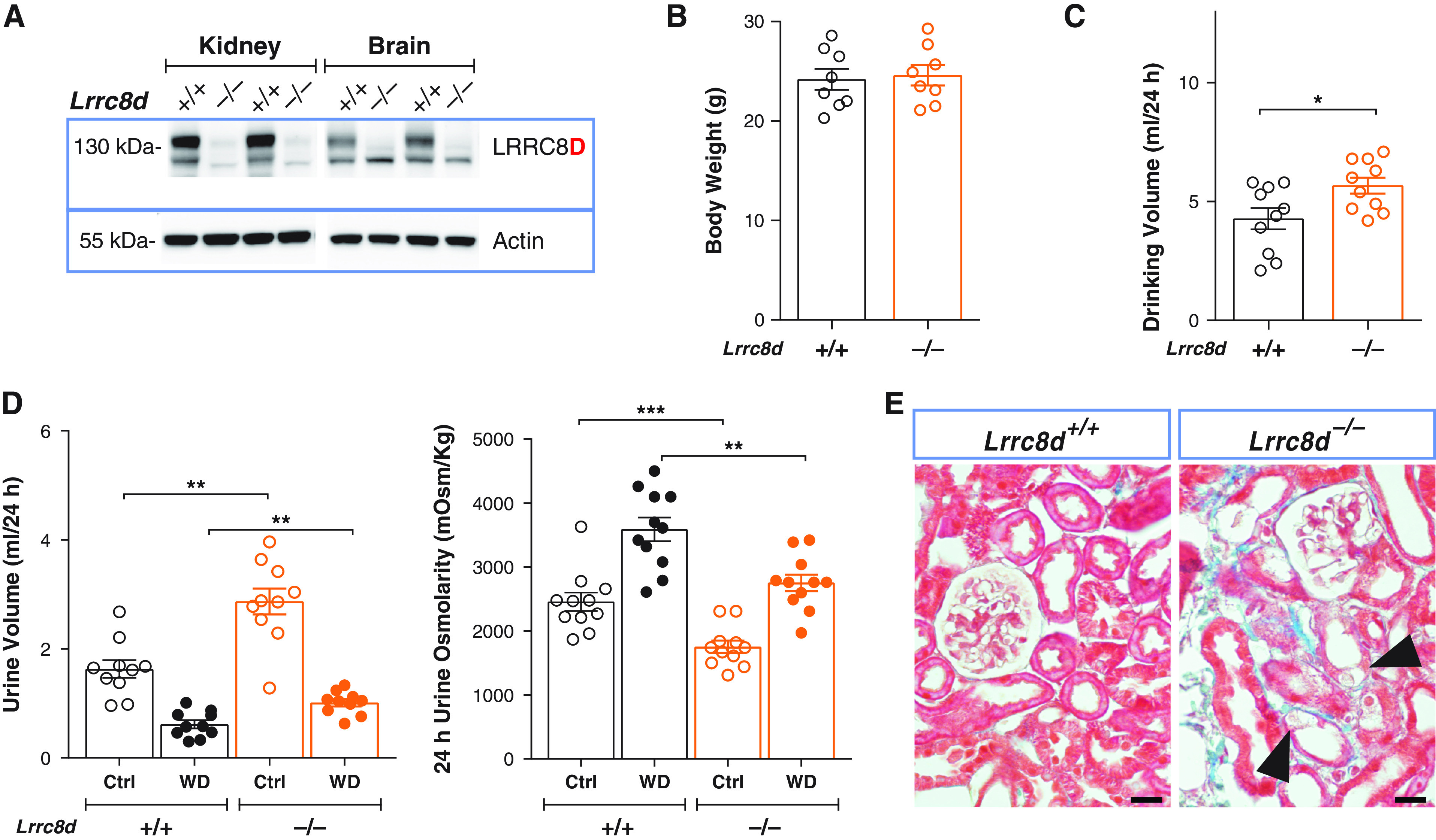
Constitutive deletion of LRRC8D recapitulates the renal phenotype of proximal tubular deletion of LRRC8A. (A) Representative Western blot analysis of LRRC8D expression in kidney and brain membrane fractions isolated from LRRC8D KO (Lrrc8d−/−) and WT mice; β-actin, loading control. (B) Body weight of 11- to 12-week-old Lrrc8d−/− and WT siblings. (C) Daily water intake of Lrrc8d−/− and WT mice. (D) Volume (left) and osmolarity (right) of 24-hour urine collected from Lrrc8d−/− and control mice, obtained with water ad libitum (Ctrl) or under 24-hour water restriction (WD). (E) Masson’s trichrome staining of kidney cortex from Lrrc8d−/− and WT mice. Mild fibrosis in Lrrc8d−/− mice (blue), tubular injury and swollen cells are indicated (arrowheads). Scale bar, 20 μm. Representative pictures of three mice per genotype. Bars, mean±SEM, **P<0.01, ***P<0.001 (Mann–Whitney U test).
