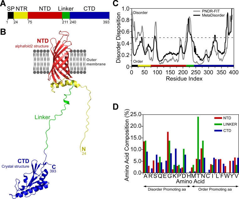
Fig 1. Structural analysis of FopA.
(A) Domain organization of FopA. The colors of the domains are maintained in the subsequent panels. SP, signal peptide; NTR, N-terminal region; NTD, N-terminal domain; CTD, C-terminal domain. (B) Alphafold2 model of FopA-NTD (red) and crystal structure of FopA-CTD (blue) solved by Michalska and co-workers (PDB ID 6U83). The unstructured linker connecting the two domains is shown in green. (C) Prediction of disordered regions in FopA by two meta disorder prediction programs PONDR-FIT [63] and MetaDisorder [60]. (D) Comparison of the composition of each amino acid in the NTD, CTD and linker. Order and disorder promoting amino acids (aa) are indicated [38].

