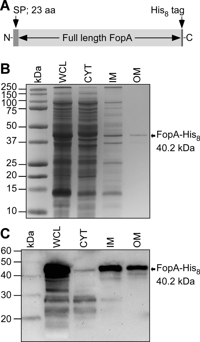
Fig 2. Subcellular localization of heterologously expressed FopA in E. coli.
(A) Schematic of the FopA expression construct. Residues 1–23 are the signal peptide (SP). (B) SDS-PAGE of E. coli subcellular fractions. WCL, whole cell lysate; CYT, cytosol; IM, inner membrane; OM, outer membrane. The bands are visualized by Coomassie stain. (C) Immunoblot using anti-His antibody of the same samples as in B.

