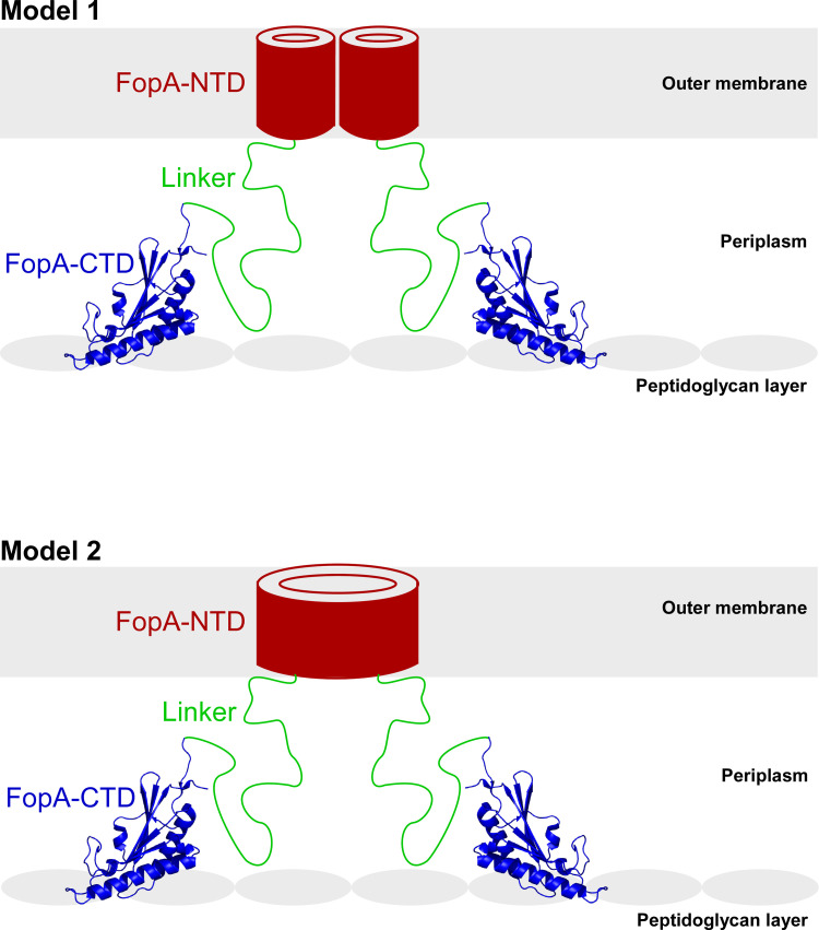
Fig 9. Proposed models of the structural organization of FopA in the outer membrane.
FopA forms a dimer. FopA-NTD (red) folds into a β-barrel and is embedded in the outer membrane, FopA-CTD (blue; PDB ID 6U83) reside in the periplasm and interacts with the peptidoglycan layer and the long flexible linker (green) tethers both domains. Model 1: the two NTDs may form two individual narrow pores; Model 2: β-strands of both NTDs may rearrange and refold into a single large pore.

