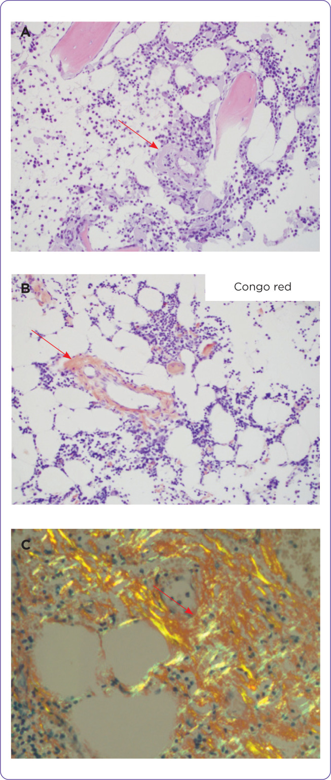
Figure 1.

(A) H & E section showing eosinophilic amorphous materials in the wall of a vessel and some in the interstitium of a bone marrow trephine biopsy. (B) Congo red stains the vessel. (C) Apple green birefringent materials in the Congo red–stained soft tissue viewed under polarized light.
