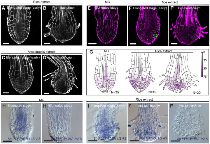
Figure 3.
Prehaustorium of S. hermonthica induced by host and nonhost root extract treatment. A–D, confocal images of Striga roots treated with rice (A and B) and Arabidopsis (C and D) roots extract. Roots were stained using Renaissance SCRI stain. Roots were treated after 24 h of germination in presence of GR24 and imaged after 4 and 12 h after treatment with root extracts. E and F′, EdU staining showing cell division in the differentiating S. hermonthica roots. G, quantification of the cell division rate during prehaustorium formation. Colored bar indicates the percentage of cell division. H–I″, RNA in situ hybridization during prehaustorium formation in longitudinal sections using ShPLETHORA1/2. H–H′ are roots germinated in MQ water. I–I″ roots treated with rice extract for 12 h after 24 h of germination. MQ, Milli-Q water, AS, Antisense, S, sense control. All scale bars: 50 µm.

