Figure 5.
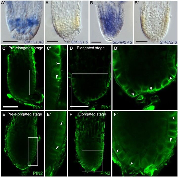
PIN protein expression and polarity in S. hermonthica during root differentiation. A–B′, RNA in situ hybridization of S. hermonthica root meristems in longitudinal sections using ShPIN1 (A–A′) and ShPIN2 (B–B′). A and B are the antisense probes and A′ and B′ are the sense control probes. C–F′, Immunolocalization using Arabidopsis thaliana PIN1 and PIN2 antibodies showing (C–D′) basal PIN1 in the epidermis. C′ and D′ are insets from C and D, respectively. N = 30. E and F′, PIN2 localization showing epidermal apical, lateral, and basal polarity. Arrows indicate the shift in PIN polarity. E′ and F′ are insets from E and F, respectively. Striga hermonthica seedlings were treated with GR24 for 24 or 48 h. N = 60. All scale bars: 50 µm.

