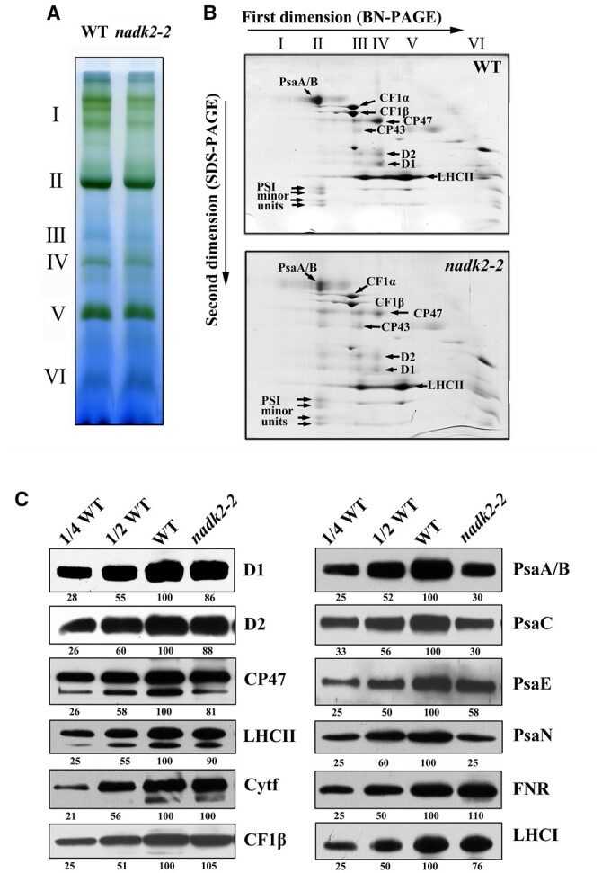
Figure 4.

Accumulation of thylakoid proteins in WT and nadk2-2 plants. A, BN gel analysis of thylakoid membrane protein complexes. Thylakoid membranes (containing 10 µg chlorophyll) from 3-week-old WT and nadk2-2 leaves were solubilized with 1% β-DM and separated on a 6%–12% gradient BN gel. Positions of distinct protein complexes are shown on the left. I, PSII supercomplex; II, PSI and dimeric PSII; III, monomeric PSII; IV, CP43-less PSII; V, trimeric LHCII and VI, free proteins. B, 2D separation of protein complexes of thylakoid membranes. After being separated on the BN gel, the protein complexes in a single lane were further separated by 15% SDS–urea–PAGE and stained with CBB. Identities of relevant proteins are indicated by arrows. C, Immunoblotting assay of thylakoid proteins in WT and nadk2-2 plants. Ten microgram of total protein extracts from 3-week-old WT and nadk2-2 leaves were separated by SDS–urea–PAGE and probed with specific antibodies. The appearing bands were scanned and analyzed using an AlphaImager 2200 Documentation and Analysis System and quantification of their relative levels are shown below the blots.
