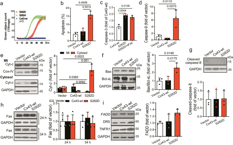
Fig. 2. Cx43-S282 mutation with aspartic acid caused cardiomyocyte apoptosis through mitochondrial apoptotic pathway.
a Time-lapse curves of NRVM death after transfection with virus carrying rat Cx43-wt, S282D or S282A gene detected by YOYO-1 probe. b–d NRVMs were transfected with virus carrying rat Cx43-wt or S282A gene, or vector for 28 h, and lysed for detection of apoptosis. Apoptotic rate was assessed by Annexin V/PI double staining and determined by a fowcytometric analysis (b). Caspase-3 (c) and caspase-9 (d) activities were detected by ELISA kits in different groups of cells. e–g The abundances of Cyt-c in mitochondria (Mt) and cytosol (e), Bax/Bcl-xL (f), and caspase-8 (g) in whole cell lysates from different groups were analyzed by Western blot. The Cyt-c fold changes in both fractions after normalized with Cox-IV or GAPDH, respectively, relative to those of control, and the fold changes in Bax, Bcl-xL and caspase-8 relative to those of Cx43-wt (after normalized with GAPDH) were thereby analyzed. h, i NRVMs were transfected with S282D or Cx43-wt gene for 24 or 34 h and the lysates were analyzed by Western blot. Fas (h), and FADD, DR5, and TNFR1 (i) were detected using specific antibody, and the fold changes in Fas and FADD relative to those of Cx43-wt (after normalized with GAPDH) were thereby analyzed. Data are expressed as mean ± SD. P values were obtained as indicated with a line between two groups in each panel using one-way ANOVA test.

