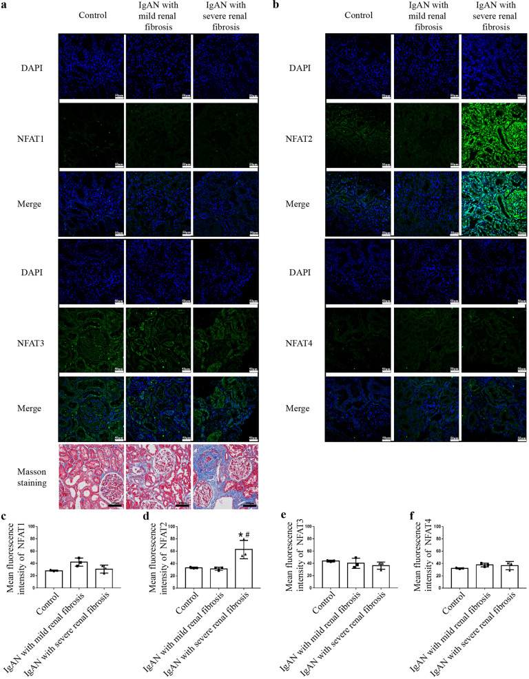
Fig. 1. NFAT2 is markedly increased in human renal tissue with severe fibrosis.
Immunostaining for NFAT1, NFAT2, NFAT3 and NFAT4 was performed on tissues from IgAN patients and adjacent normal tissues from renal cell carcinoma patients. Representative confocal images showed higher expression and more nuclear localization of NFAT2 in the renal tubules, interstitium and glomerulus in IgAN patients with severe renal fibrosis (n = 3) than in IgAN patients with mild renal fibrosis or in adjacent normal renal tissue from renal cell carcinoma patients (n = 3) (Scale bars = 50 μm). Masson staining was used to assess kidney fibrosis (scale bars = 100 μm). a Immunostaining for NFAT1 and NFAT3. b Immunostaining for NFAT2 and NFAT4. c–f Quantitative analysis for the immunofluorescence of NFAT1, NFAT2, NFAT3 and NFAT4. *P < 0.05 vs. Control; #P < 0.05 vs. IgAN with mild renal fibrosis. NFAT1 nuclear factor of activated T cells 1, NFAT2 nuclear factor of activated T cells 2, NFAT3 nuclear factor of activated T cells 3, NFAT4 nuclear factor of activated T cells 4.

