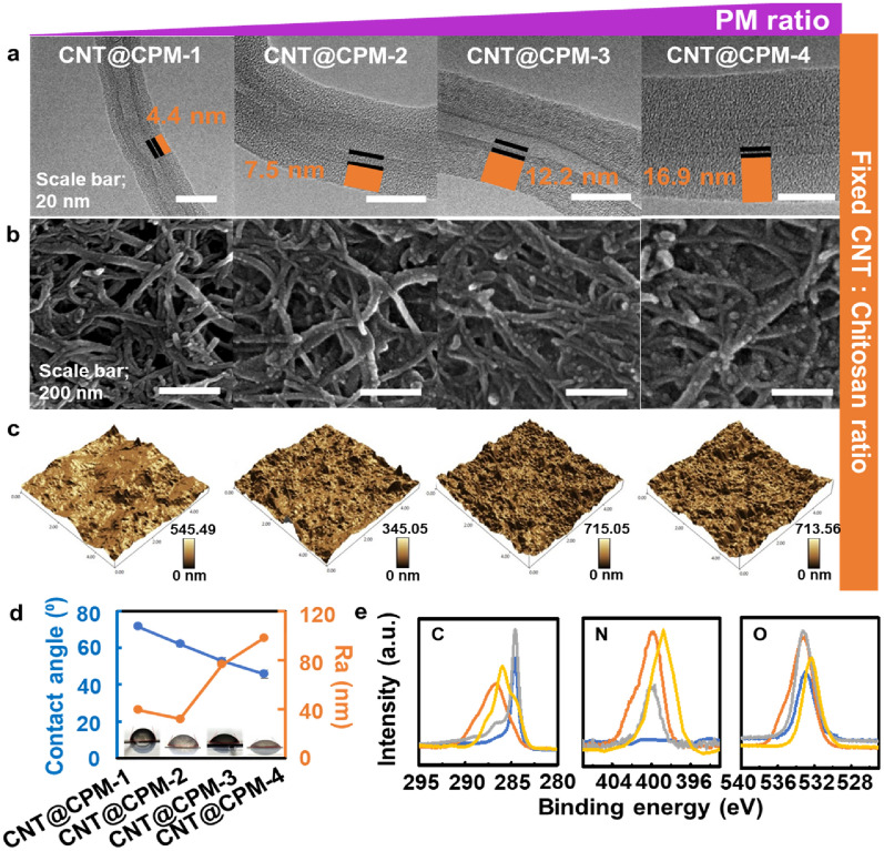
Fig. 2.
The morphology and physicochemical properties of nanocomposites formed at different PM concentrations. a HR-TEM images of shell thickness ranged from 4.4 to 17.4 nm depending on the PM ratio. b FE-SEM images of CNT@CPM; CPM shell thickness became thicker. c AFM surface images of CNT@CPM, exhibiting a highly rough nanoto-pographical surface, d with a roughness ranged from 32.1 nm to 98.2 nm as the ratio of PM increased, and contact angles; hydrophilicity varies depending on concentration of PM. e XPS scanned revealing that the self-assembled CNT@CPM had clear shift of binding energy

