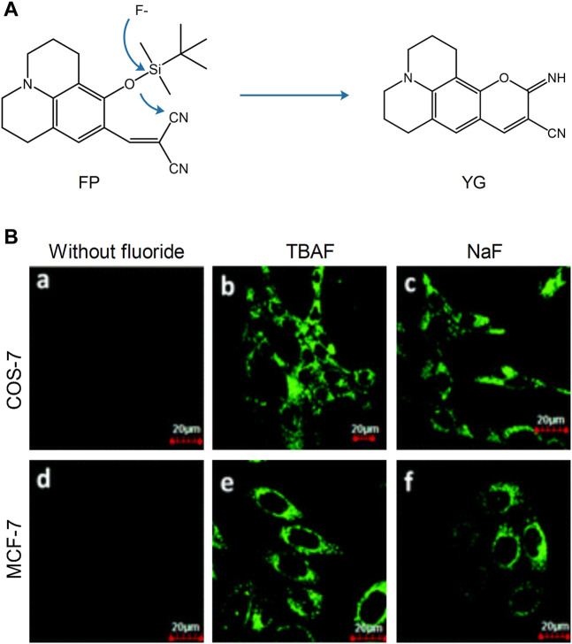
FIGURE 5.
The reaction mechanism and fluorescence imaging of probe FP (Shiling et al., 2014). (A) Proposed reaction mechanism of FP. (B) Fluorescence imaging of COS-7 and MCF-7 cells incubated with probe FP (2.5 μM) before (a and d) and after (b, c, e, and f) being treated with TBAF, NaF (100 μM).

