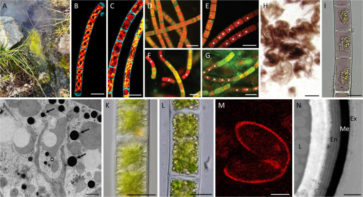
FIGURE 2.
Effects and adaptation mechanisms of Zygnematophyceae to high radiation and temperature extremes. (A) Zygnematophyceae mat in semi-terrestrial habitat; (B) confocal micrographs of Spirogyra sp. maintained at 22°C; (C) confocal micrographs of Spirogyra sp. maintained at 37°C with altered plastids; (D–G) Zygnema sp. stained with 0.1% Auramine O, dead cells marked with an asterisk; (D) young culture (–2°C); (E) young culture (–10°C); (F) pre-akinetes (–20°C); (G) pre-akinetes (–70°C); (H) purple Zygogonium ericetorum filaments; (I) cells of Z. ericetorum with purple pigment in the vacuoles; (J) electron micrograph of Zygnema sp. exposed to PAR + UV-A showing electron-dense particles (arrows); (K) young Zygnema sp. cells after PAR + UV-A + UV-B (PAB) treatment; (L) pre-akinetes after PAB treatment; (M) RAMAN imaging of Spirogyra mirabilis zygospore showing aromatics in cell wall; (N) transmission electron micrograph of Spirogyra mirabilis zygospore cell wall showing three-layered structure and electron-dense middle layer; Abbreviations: En, endospore; Ex, exospore; Me, mesospore; and L, lipid. Scale bars (B,C) 100 μm; (D–G) 40 μm; (H) 1 cm; (I,K,L) 20 μm; (J) 1 μm; (M) 15 μm; (N) 1 μm. (B,C) Reprinted from de Vries et al. (2020); (D–G) Reprinted from Trumhová et al. (2019); (H,I) Reprinted from Aigner et al. (2013); (J) Reprinted from Pichrtová et al. (2013); (K,L) Reprinted from Holzinger et al. (2018); and (M,N) Reprinted from Permann et al. (2021a). All reprinted material was published under CC-BY license and is copyrighted by the authors.

