
FIGURE 2.
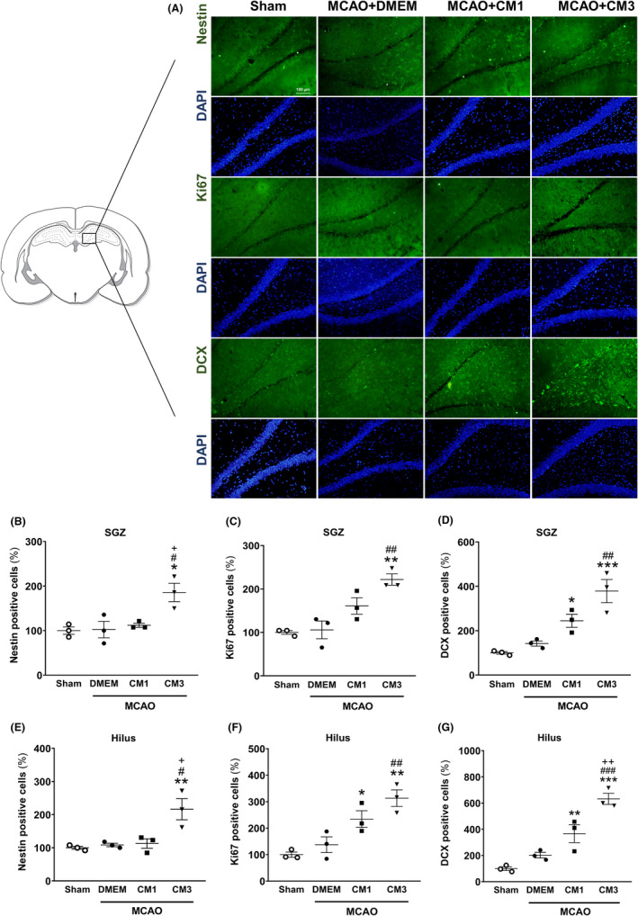
Effect of hESC‐MSC‐CM on protein expression of neurogenesis markers. (A) Representative micrographs of immunofluorescence staining of Nestin, Ki67, and DCX. Cell nuclei were counterstained with DAPI. Scale bar: 100 μm. The percentage of (B) Nestin, (C) Ki67, and (D) DCX‐ positive cells relative to sham in the SGZ. The percentage of (E) Nestin, (F) Ki67, and (G) DCX‐ positive cells relative to sham in the hilus. Data are reported as the mean ± SEM (n = 3); the differences between groups were determined by ANOVA followed by Tukey test. *p < 0.05, **p < 0.01, and ***p < 0.001 vs. Sham, # p < 0.05, ## p < 0.01 and ### p < 0.001 vs. MCAO+DMEM, + p < 0.05 and ++ p < 0.01 vs. CM1. CM, conditioned medium; DCX, Doublecortin; DMEM, Dulbecco's modified eagle's medium; MCAO, middle cerebral artery occlusion; SGZ, subgranular zone