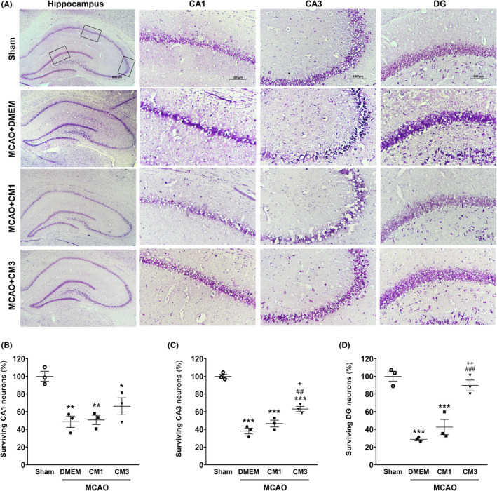
FIGURE 7.

Effect of hESC‐MSC‐CM on neuronal survival. (A) Representative micrographs of Nissl‐stained sections in the CA1, CA3, and DG hippocampal subfields. Scale bar: 100 μm. (n = 3). The percentage of surviving neurons in (B) CA1, (C) CA2, and (D) DG in the hippocampus. *p < 0.05, **p < 0.01 and ***p < 0.001 vs. Sham, ## p < 0.01 and ### p < 0.001 vs. MCAO+DMEM, + p < 0.05 and ++ p < 0.01 vs. CM1. CM, conditioned medium; DMEM, Dulbecco's modified eagle's medium; MCAO, middle cerebral artery occlusion; DG, dentate gyrus
