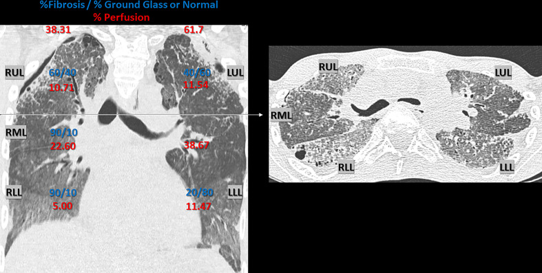
Figure 1.
Chest CT Scan 1 year pre-lung transplant. Chest CT Scan 1 year pre-lung transplant (2016), with representative coronal and axial section, demonstrating interstitial lung disease, with parenchymal fibrosis and ground glass opacities, due to alveolar lipoproteinosis caused by hereditary pulmonary alveolar proteinosis. Radiologic signs of irreversible fibrosis included thickening of intra- and interlobar septae (reticulation), traction bronchiectasis and honeycombing. Architectural distortion was more prominent in the anterior en apical lung fields compared to the postero-basal lung fields. A secondary spontaneous partial pneumothorax was present, situated bilateral apico-lateral (dehiscence: left side 8 mm and right side 5 mm). An estimate of the ratio (%) fibrosis vs. ground glass opacification or normal appearing lung parenchyma per lobe is given in blue (visual scoring by a skilled chest radiologist with expertise in pulmonary fibrosis). The respective perfusion (%) of each lung and individual lobe is given in red, illustrating regional differences due to hypoxic vasoconstriction. Perfusion was measured using technetium-99m radioisotope (122.0 MBq, 3.30 mCI) and Pulmonics software. RUL, right upper lobe; RML, right middle lobe; RLL, right lower lobe; LUL, left upper lobe; LLL, left lower lobe.

