Fig. 6.
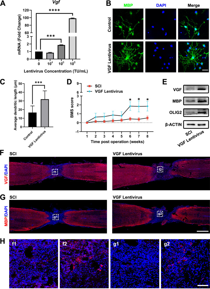
Overexpression of VGF promoted oligodendrogenesis in vitro and in vivo. A qPCR detection of Vgf after VGF overexpression in cells using various doses of lentivirus. B Immunostaining of MBP (green) under treatment with 108 TU/mL VGF lentivirus. Nuclei were stained with DAPI (blue). Scale bar, 10 μm. C Quantification of the average dendritic length. D BMS open-field walking scores of the mice treated with VGF lentivirus for 8 weeks. *p < 0.05 compared to the SCI group. E Western blot detection of VGF, MBP, and OLIG2 expression in the injured spinal cord after VGF lentivirus treatment. F, G Staining of VGF and MBP in the lesioned spinal cord. Scale bar, 1000 μm. H Amplified images of highlighted regions at the lesion site of mice. Scale bar, 100 μm

