Fig. 1.
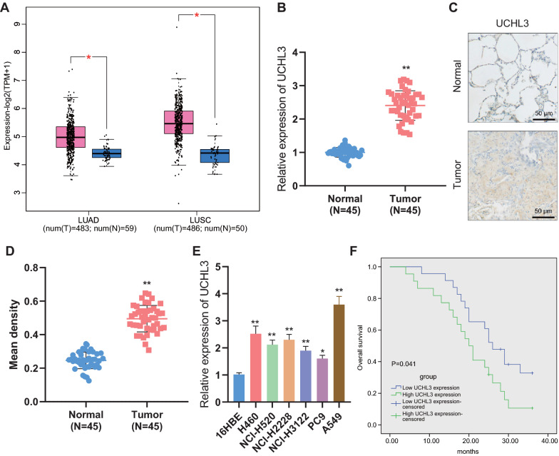
UCHL3 expression was elevated in NSCLC tissues and cells. A, The expression of UCHL3 in TCGA-LUAD and TCGA-LUSC datasets from the GEPIA database (T represents tumor tissue, displayed as a red column; N represents normal tissue, displayed as a blue column). B, qRT-PCR measurement of UCHL3 expression in tumor tissues and adjacent normal tissues from NSCLC patients (n = 45). C, Positive expression of UCHL3 protein in tumor tissues and adjacent normal tissues from NSCLC patients (n = 45) detected by Immunohistochemistry. D, Quantitative results of panel C. E, qRT-PCR measurement of UCHL3 expression in NSCLC cell lines (H460, NCI-H2228, NCI-H3122, PC9 and A549) and human bronchial epithelial cell line 16HBE. F, Correlation between the expression of UCHL3 and the survival rate of NSCLC patients analyzed by the Kaplan–Meier method. * p < 0.05, ** p < 0.01 versus adjacent normal tissues or 16HBE cells. Each cellular experiment was repeated 3 times

