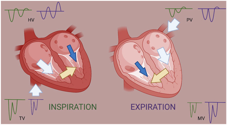
Fig. 3.
Schematic representation of ventricular interdependence. During inspiration (green color), the blood return to the right side (white arrow) increases leading to higher tricuspid valve inflow velocities (green TV Doppler) but thickened pericardium prevents outward expansion of right ventricle leading to shifting of septum (yellow arrow) towards left side, thereby contributing to decreased blood flow on left side (blue arrow) and lower mitral inflow velocities (green MV Doppler). During expiration (purple color), the inflow to left ventricle increases (white arrow) depicted by increased mitral inflow velocity (purple MV Doppler), causing shifting of septum (yellow arrow) towards right side contributing to decreased blood flow on right side (blue arrow) and lower tricuspid inflow velocities (purple TV Doppler) (Created with BioRender.com)

