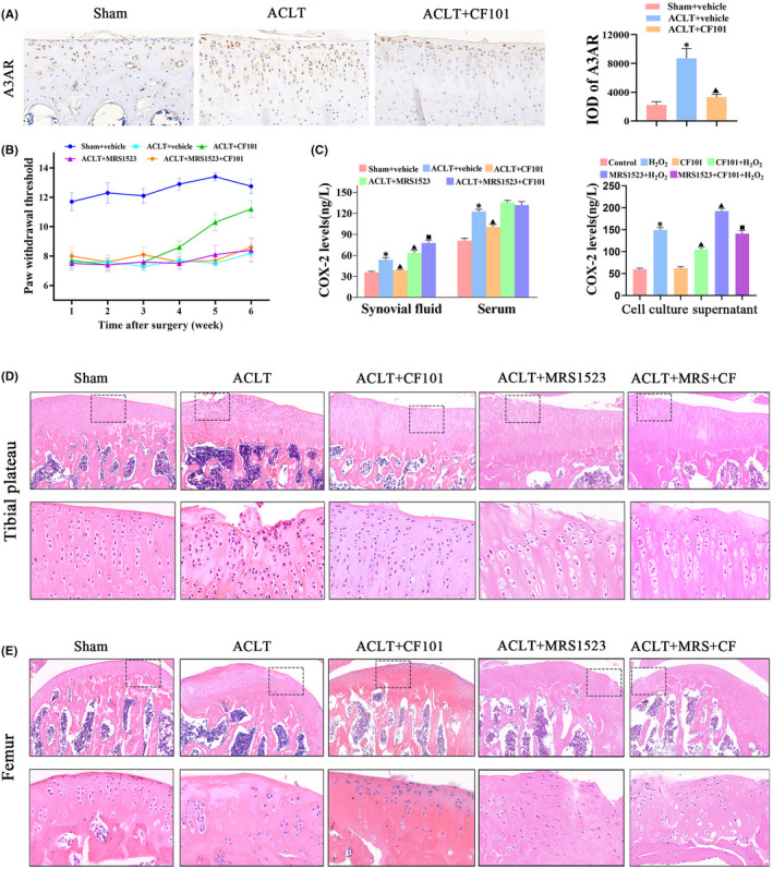
FIGURE 1.

A3AR activation by CF101 ameliorated OA‐related pain and protected the structure of cartilage. (A) IHC analysis of A3AR in tibial plateau of right knee joints (n = 3, the positive stained chondrocytes in three central regions of articular cartilage were counted using Image Pro Plus version 6.0 software), Original magnification × 200. (B) Effect of CF101 on von Frey hair withdrawal threshold in ACLT‐induced rats (n = 12). (C) ELISA assay of COX‐2 in synovial fluid, serum and cell culture supernatant (n = 6). (D) The pictures above are representative images of cartilage in tibial plateau (right knees of rats) stained by hemotoxylin and eosin (HE), Original magnification × 100. The picture below is expansion of the region occupied by articular cartilage (n = 6), Original magnification × 400. (E) The pictures above are representative images of cartilage in femur (right knees of rats) stained by hemotoxylin and eosin (HE), Original magnification × 100. The picture below is expansion of the region occupied by articular cartilage (n = 6), Original magnification × 400. Values are the mean ± SEM; *p < 0.05 vs. Sham group. ▲ p < 0.05, vs. ACLT group. ■ p < 0.05, vs. ACLT + CF101 group
