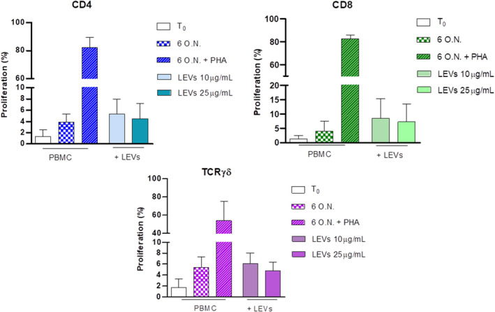
FIGURE 4.

Influence of LEVs on CD4, CD8 and γδ T cell proliferation was evaluated after 6 days at two different concentrations, 10 and 25 μg/ml. PBMCs stimulated with PHA were used as the positive control (striped bar). The percentage of proliferated cells was determined using Tag‐it Violet
