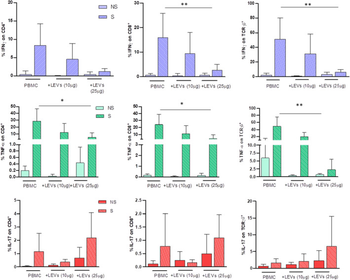
FIGURE 6.

Pro‐inflammatory cytokine expression on CD4, CD8 and γδ T cells was evaluated by flow cytometry. The effect of 10 and 25 μg/ml of LEVs was compared with untreated PBMC. Every condition was performed with (S, striped bar) or w/o (NS, full/coloured bar) ionomycin and PMA. Statistical significance was determined by Kruskal–Wallis. p < 0.05 (*), p < 0.01 (**)
